Sign Up
School Account Sign Up
Lesson ID: 12165
Zoom into the tiny world of electron microscopes and learn when scientists use SEM, TEM, or a classroom microscope to solve big questions!


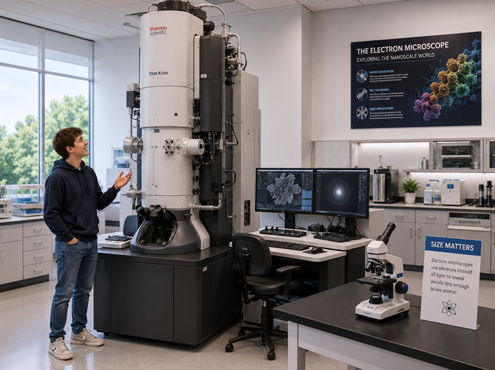
A Microscope the Size of a Vending Machine?
Imagine trying to take a selfie with something so small that a regular microscope says, “Nope. Too tiny.”
A red blood cell? Tiny. A bacterium? Tinier. A virus? Even tinier.
Some cell parts and molecules are so small that visible light cannot show them clearly.
That is where electron microscopes come in.
A regular classroom microscope, called a compound light microscope, uses visible light and glass lenses to magnify small objects. It works well for viewing things such as onion cells, pond water organisms, and thin slices of plant or animal tissue.
An electron microscope uses a beam of electrons instead of light. Electrons are tiny negatively charged particles found in atoms. Because electrons can act like waves with much shorter wavelengths than visible light, electron microscopes can reveal much smaller details than light microscopes can.
In other words, a light microscope gives you a close look. An electron microscope gives you a “whoa, that was hiding there?” look.
Why Electrons Show More Detail
To understand why electron microscopes are so powerful, think about trying to draw with two different markers.
A thick marker works for big shapes, but it cannot show tiny details. A fine-tip pen can draw sharper lines and smaller patterns.
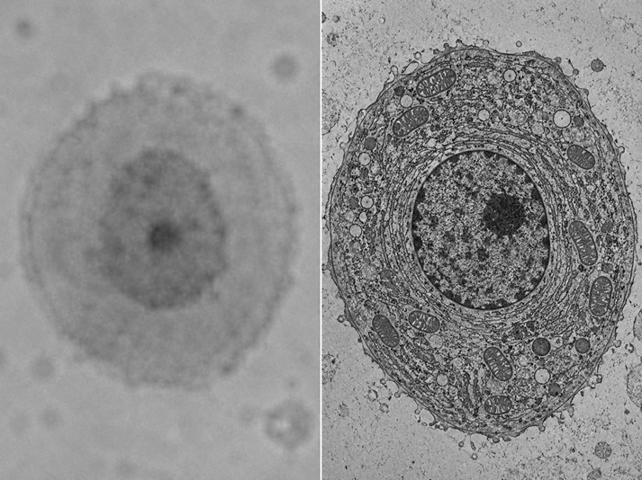
Visible light works a little like the thick marker. Its wavelength limits how much detail a light microscope can show.
Electrons have much shorter wavelengths, so they can reveal smaller details. This sharper view is called better resolution.
Resolution means how clearly a microscope can show two separate points as separate. Magnification means how much bigger an image appears.
Both matter, but resolution is the real star. A blurry image that is made bigger is still blurry. Electron microscopes help scientists see tiny structures more clearly, not just larger.
Electron microscopes can help scientists study things such as cells, organelles, bacteria, viruses, proteins, materials, minerals, and the surfaces of tiny objects.
Scientists use them in medicine, biology, chemistry, engineering, environmental science, materials science, and forensic science.
How an Electron Microscope Works
An electron microscope does not look like the microscope in a classroom. It is usually large, expensive, and found in a lab. It also needs special conditions to work.
Here is the basic idea:
An electron gun produces a beam of electrons.
Electromagnetic coils focus and steer the electron beam. These coils act like lenses, but they use magnetism instead of glass.
The electron beam interacts with the sample.
Detectors collect information from the electrons.
A computer turns that information into an image.
Electron microscopes use electromagnetic coils because electrons cannot pass through glass lenses the way light does. The microscope must control the electrons with magnetic fields.
Most electron microscopes also work in a vacuum. A vacuum removes most air particles from the microscope’s chamber.
This matters because air particles could bump into the electron beam and scatter it before it reaches the sample.
Samples also need special preparation. Scientists may dry, freeze, slice, stain, coat, or preserve a sample before viewing it.
Because of that preparation, traditional electron microscopes usually do not show living things moving in real time. Instead, they give scientists a detailed snapshot of a sample at one moment.
That snapshot can still reveal a lot. One image can show the bumps on a pollen grain, the shape of bacteria, the inside of a cell, the structure of a virus, or the tiny crystals inside a mineral.
Two Main Types: SEM and TEM
Scientists choose a microscope based on the question they want to answer. Two main types of electron microscopes are scanning electron microscopes and transmission electron microscopes.
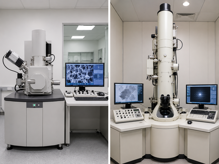
Scanning Electron Microscope: SEM
A scanning electron microscope, or SEM, shows the surface of a sample.
The SEM scans a focused beam of electrons across the outside of an object. When the electrons hit the surface, they cause other electrons and signals to bounce away from the sample. Detectors collect those signals and build an image.
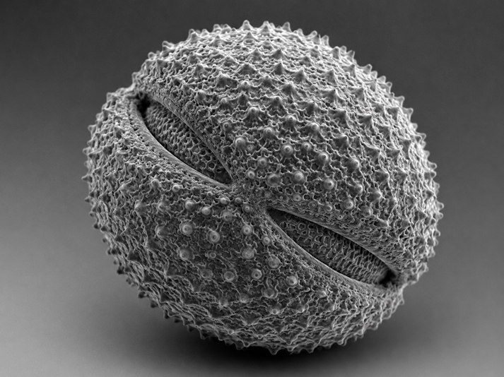
SEM images often look three-dimensional because they show surface shape and texture. An SEM works well when scientists want to study the outside of something.
A scientist might use an SEM to study:
the surface of an insect’s eye
the texture of a leaf
the shape of bacteria
the structure of a snowflake
the surface of a metal, rock, or plastic
tiny clues in forensic evidence, such as hair, fibers, or tool marks
SEM images are usually black-and-white when produced by the microscope. Sometimes artists or scientists add color later to help people understand different parts of the image.
Adding color can make an image easier to study, but it does not show the object’s natural color.

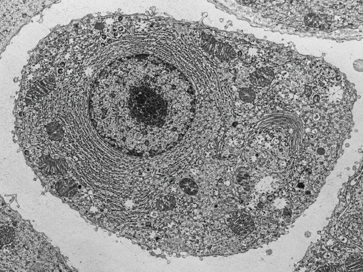
Transmission Electron Microscope: TEM
A transmission electron microscope, or TEM, shows the inside structure of a very thin sample.
The TEM sends electrons through the sample. Some electrons pass through, and others are blocked or scattered by the sample’s structures. The microscope uses that information to create an image.
Because the electrons must pass through the sample, the sample has to be extremely thin. Scientists often slice cells or tissues into ultra-thin sections before placing them in a TEM.
A TEM works well when scientists want to study internal details.
A scientist might use a TEM to study:
parts inside a cell
organelles, such as mitochondria
viruses
protein structures
thin tissue samples
cell membranes
tiny structures that help cells work
TEM images can show amazing detail, but they usually do not look three-dimensional in the same way SEM images do. A TEM is more like looking through a thin slice. An SEM is more like looking at the outside surface.
SEM or TEM: Which One Should a Scientist Choose?
The best microscope depends on the question.
If a scientist asks, “What does the outside look like?” an SEM is usually the better choice.
If a scientist asks, “What is inside?” a TEM is usually the better choice.
For example, imagine a scientist studies a mysterious seed.
If the scientist wants to see the seed’s surface texture, cracks, ridges, or tiny hairs, the SEM would be useful.
If the scientist wants to see thin slices of cells inside the seed, the TEM would be useful.
Now, imagine a scientist studies a virus.
A TEM may help reveal the virus’s structure because viruses are extremely small and lack large external surfaces like those of insects or seeds. Scientists may also use other specialized electron microscopy methods, depending on what they need to learn.
The microscope is not “better” by itself. The better microscope is the one that answers the question.
More Than Pretty Pictures
Electron microscope images can look strange, beautiful, or even a little creepy. A dust mite may look like a movie monster. Pollen may look like a spiky space pod. A tiny crystal may look like a mountain range.
These images are more than cool science art. Scientists use them to solve real problems.
Electron microscopes can help researchers understand how cells function, how diseases affect tissues, how strong materials fail, how minerals form, how bacteria attach to surfaces, and how tiny structures influence everyday products.
Engineers may use electron microscopes to inspect computer chips, medical devices, metals, or new materials. Forensic scientists may use them to compare tiny pieces of evidence. Medical researchers may use them to study cells, viruses, and proteins.
The big idea is simple: better tools help scientists ask better questions. As microscopes improved, scientists saw details they could not study before. Those details opened the door to new discoveries.
What You Need to Remember
An electron microscope uses electrons instead of visible light.
Electrons have much shorter wavelengths than visible light, so electron microscopes can show much smaller details.
Resolution means how clearly a microscope shows separate details.
Electron microscopes use electromagnetic coils instead of glass lenses.
Most electron microscopes require a vacuum and special sample preparation.
A scanning electron microscope, or SEM, shows surface details.
A transmission electron microscope, or TEM, shows internal details in thin samples.
Scientists choose SEM or TEM based on what they need to find out.
Now that you know how electron microscopes work and how SEM and TEM images differ, you are ready to practice choosing the right microscope for different scientific questions.

Supplies


