Sign Up
School Account Sign Up

Lesson ID: 11643
Discover how viruses invade cells, take over the machinery, and burst out in a dramatic finish—one cell at a time!




Imagine a tiny invader so small you’d need an electron microscope to see it—but so effective, it can turn a healthy cell into a virus factory and blow it apart in a matter of minutes.
This is no horror movie villain—it’s the lytic cycle in action, a viral replication strategy that ends with destruction.
How Viruses Reproduce: The Lytic Cycle
Viruses are microscopic particles made of genetic material (DNA or RNA) enclosed in a protein shell called a capsid. Some viruses also have an envelope made from the host cell’s own membrane.
But unlike living cells, viruses don’t eat, grow, or reproduce on their own. They’re obligate intracellular parasites—they must hijack a living cell to make more copies of themselves.
One of the fastest and most destructive ways they do this is through the lytic cycle. Follow a bacteriophage—a virus that infects bacteria—as it goes through this dramatic process.
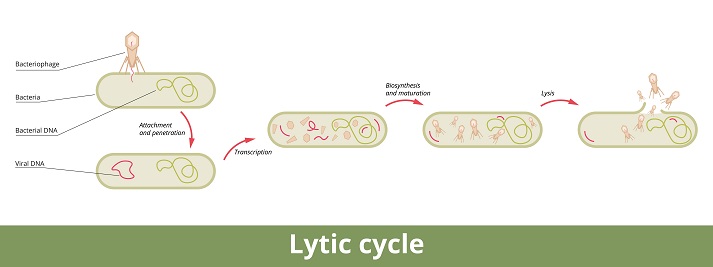
Step 1: Attachment
The virus searches for a specific host cell with matching receptors. Think of it like a key looking for a lock.
Tail fibers on the virus recognize and bind to proteins on the surface of the host. In this case, the bacteriophage T4 locks onto an E. coli bacterium.
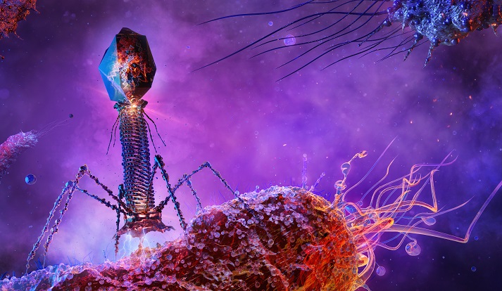
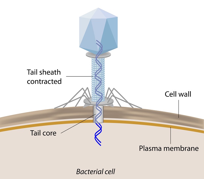
Step 2: Entry
Once attached, the phage’s tail sheath contracts like a syringe. A powerful protein called gp5, acting as a molecular drill and enzyme, punctures the bacterial cell wall and membrane.
The viral DNA is then injected into the host’s cytoplasm while the empty capsid stays outside.
Step 3: Replication & Gene Expression
Inside the host, the virus takes control of the cell’s machinery. The host’s DNA is broken down to free up building blocks.
The viral genome begins to replicate, using both host enzymes and its own specialized proteins. Viral RNA is transcribed and translated into proteins, including capsids, tail structures, and enzymes needed for assembly and escape.
Step 4: Assembly
New viruses are built inside the cell. Capsids form and are filled with replicated DNA. Tails and fibers are attached.
Like an assembly line, dozens of complete viral particles are rapidly put together, right inside the host.
Step 5: Release
Once enough viruses are assembled, the cell is destroyed.
Viral proteins called holins punch holes in the bacterial membrane, and enzymes called lysozymes break down the cell wall. The cell bursts open—a process called lysis—releasing a swarm of new viruses into the environment, ready to repeat the cycle.
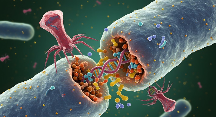
This fast and furious cycle is why lytic viruses are so dangerous—they don’t wait around. They get in, take over, explode out, and move on.
Watch It Happen
Before moving on, watch this short animation that brings the lytic cycle to life. You’ll see each stage unfold—from viral attachment to cell destruction—in a real-time example of how efficient and destructive this process can be.

Keep Going
Now that you’ve seen how a virus turns a host cell into a factory, it’s time to test your knowledge and try your hand at breaking down the process yourself.
Head to the Got It? section.

Supplies
Additional Resources


 FAVORITE
 Like
Like